Momo pregnancy management

Disclaimer: The following post summarizes what I learned to expect for managing my monoamniotic pregnancy. THIS POST IS NOT PROVIDING MEDICAL ADVICE. Everyone’s pregnancy is different. Consult your obstetrician for medical advice; not my blog!

Cord entanglement
In-patient vs. out-patient monitoring
Gestation beyond 34 weeks
Caesarean vs. vaginal birth

By my 16th week of pregnancy, I understood that my twins were likely monoamniotic: the rarest and riskiest twin type to carry. The two weeks between my diagnosis at the perinatologist clinic and my subsequent meeting with my obstetrician were grueling. I needed assurances that my twins were going to be okay; I craved a game plan for how we were going to keep me and my girls safe (I found out my twins were identical girls in the first trimester from a blood test). I kept my mind preoccupied with research. I searched for anecdotes, and I searched for statistics. It was hard to feel optimistic based on the information readily available to anyone online, but the scientific literature published within the last five or so years had a very different tone.

How the internet makes a bad first impression

Like any complicated medical condition, initial internet searches of monoamniotic twin pregnancy reveal depressing statistics.

From obgyn-care.net: “To have an idea about the increase of vital risk, you should know that twins growing in the same sac (monochorionic, monoamniotic) have a survival rate of only 50%!”

From monoamniotic.org: “Among the couples who…were truly diagnosed as monoamniotic, 60% have had two healthy babies.”

Not very uplifting.

Maybe reading anecdotal experiences would put my mind at ease? Fortunately, there were plenty of success stories spanning five decades to assure me that monoamniotic twins were not fated to die in-utero. But some people’s stories from just within the last decade exposed the disturbing beliefs of some physicians about monoamniotic twin pregnancies. A woman pregnant with momo twins in 2008 wrote that her physician informed her that “there wasn’t much of a chance of survival” and that she could “continue…a hellacious pregnancy and still possibly lose both babies or terminate the pregnancy.” She chose to terminate and now lives with regret. Upon diagnosis of monoamniotic twins, one man in 2011 wrote that his MFM doctor offered him and his wife two options: “terminate the pregnancy, or continue.” “In many cases, we would recommend for the couple to terminate this kind of pregnancy…” the man quotes his physician.

Why were those physicians so dire? Of course without more information it is difficult to understand the complete circumstances of those pregnancies. There are many ways a monoamniotic pregnancy can be complicated with a tenuous outcome. Sudden death from cord constriction is particularly concerning and commonly cited when discussing the perils of a monoamniotic twin pregnancy. But does it justify threatening an expecting family with loss?

Cord entanglement in monoamniotic twins

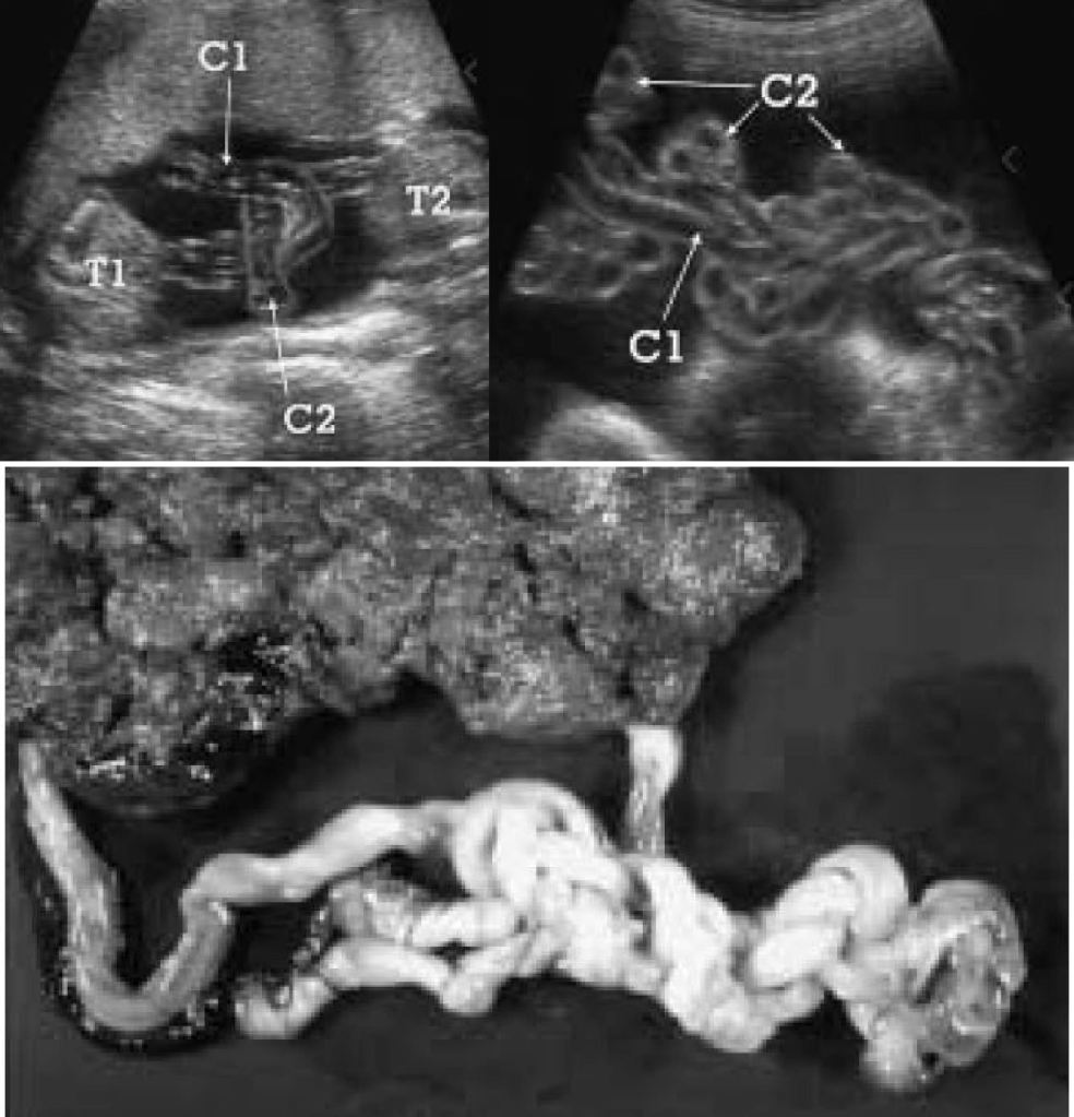
The short answer is no, most monoamniotic twins are not going to spontaneously die from cord entanglement. The truth is, entanglement of the umbilical cord is expected in 100% of momo gestations (Dias et al., 2010)(Fig. 2). Constriction of the cords, not just entanglement, is the real threat to momos, but not all cords become so tightly knotted. Most prenatal mortality before 20 weeks results from twin-reversed arterial perfusion (TRAP), discordant anomalies, spontaneous miscarriage from developmental anomalies, conjoined twinning, and complications from prematurity, but not from cord entanglement itself (Dias et al., 2010; Rossi and Perfumo, 2013). Size discordance in monochorionic twins is a symptom of twin-to-twin transfusion syndrome (TTTS). If left untreated, it can lead to intrauterine death of both twins. The silver lining from expecting momo twins is that TTTS is five times less common in momos than in di-mos. This is likely due to the arterial connections present in the placenta of momos, but not typically in di-mos (Umer et al, 2003; Hack et al., 2009). However, congenital defects are more common in momos (Myrianthopoulos, 1975). So long as these congenital defects are not detected in the 20 week anatomy scan and the succeeding echocardiogram around 24 weeks, a family expecting momos can breathe a sigh of relief, for the outcome of developmentally normal monoamniotic twins after 20 weeks is likely to be positive. If the cords are twisted or even knotted to some degree, do not panic; it is not an automatic death sentence. However, do consider that as the fetuses grow in size, so too does the diameter of their cords. Therefore, the threat of any tangled and knotted cords becoming compressed occurs later in gestation. That is part of the reasoning behind delivering momo twins no later than 32-34 weeks. Fortunately, the number of spontaneous losses of momo twins has decreased since the improvement of monitoring practices toward a more intensive monitoring schedule (Roqué et al., 2009; Rossi and Perfumo, 2013).

Figure 2. Progression of tangled umbilical cords in a monoamniotic twin pair. Top left: Ultrasound image of entangled cords at 14 weeks (Tongsong and Chanprapaph, 1999: fig. 1). Top right: Ultrasound image of the same entangled cords at 26 weeks (Tongsong and Chanprapaph, 1999: fig. 4). Bottom: Photograph of same knotted and twisted cords after delivery at 34 weeks (Tongsong and Chanprapaph, 1999: fig. 5). Twins survived and were healthy. C = cord, T = twin.

In-patient vs. out-patient monitoring

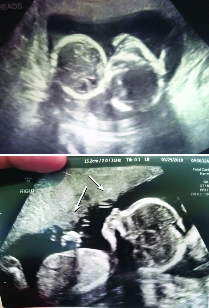
What does intensive monitoring mean when you are expecting momos? At the very least, a mother of momos can expect non-stress testing (NST) every two to three days, coming and going to her monitoring appointments at her leisure. Depending on her hospital and obstetrician policies, she can otherwise expect NSTs two to three times a day with ultrasounds twice a week (Fig. 3). Such regular monitoring would require the mother to go inpatient. That means living in the hospital without discharge until the babies are born and the mother is fit to go home.

Figure 3. Non-stress test output from one of my inpatient monitoring sessions. The blue and pink lines represent the heart rate of each twin, while mine is in green. Heart decelerations and accelerations are to be expected, but deep dips like the one seen on the blue line to the left of the screen were cause for concern if they persisted for a minimum of ten minutes and were not followed by a spike in the heartrate. One day during one of my monitoring sessions, a dip lasted seven minutes and I was moved from Antepartum to Labor & Delivery, and prepped for surgery. I was able to keep the twins in for another three weeks, but such dips would extend my monitoring sessions by 20 minutes to an hour, minimum. My monitoring schedule was seldom adhered to, so outpatient monitoring would have been more trouble than it was worth as I could not rely on any particular in-and-out time.

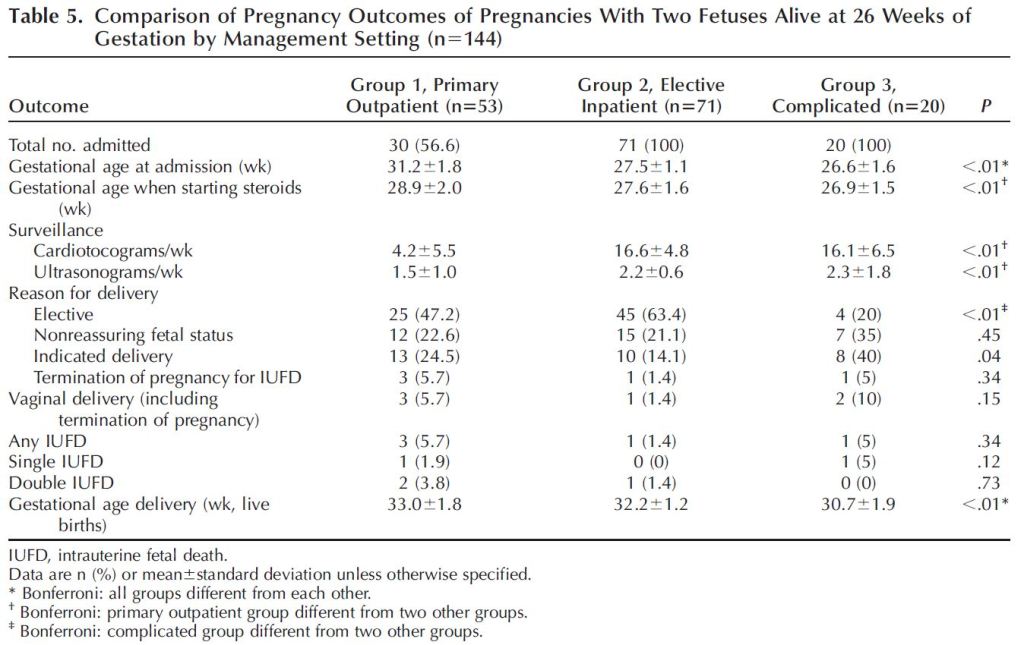
One can imagine the quality of life for the mother and her family varies significantly between the outpatient and inpatient options. Outpatient monitoring would allow one to be home with her husband and any children she may already have. Additionally, the financial burden of an extended hospital stay would be significantly reduced. Further, a mother living inpatient in the hospital may reduce her activity enough to put herself at risk of a thromboembolism, a dangerous blood clot. However, remaining inpatient would allow for more frequent monitoring and access to immediate medical attention in the event of an emergency. Further, for those families who live outside of major cities and a good distance from hospitals that can manage monoamniotic pregnancies, even twice-weekly visits may be a greater burden than just living in the hospital. Which option should you choose? You honestly may not have much of a say in how you are monitored if you wish to keep your obstetrician. One of the MFM specialists I visited favored outpatient monitoring over inpatient monitoring for some of the reasons discussed above. But my obstetrician had the final say and she was very adamant that I go inpatient. Inpatient monitoring was supported in a 2005 study that reported no intrauterine death in the cohort of patients who went inpatient, vs. perinatal loss in patients who were monitored as outpatient (Heyborne et al., 2005). The study also reported higher neonatal birthweight, older gestational age at delivery, and improved newborn survival among babies born from mothers who electively admitted themselves for inpatient monitoring (Fig. 4). One caveat to their results was that mothers who were eventually admitted for inpatient monitoring due to signs of complications (arising from, for example, TTTS) were grouped with the outpatient cohort, which could have biased results since the outpatient cohort was populated by complicated monoamniotic gestations.

Figure 4. Table 5 from Heyborne et al., 2005, summarizing statistics and outcomes of patients who electively went inpatient vs. patients who remained outpatient until there were signs of complications. The authors found significant differences in fetal survival rate, gestational age at delivery, and birth weight between the outpatient and inpatient cohort, with results favoring inpatient monitoring. However, it is important to note that patients who were eventually admitted for inpatient monitoring due to pre-existing complications were combined with the outpatient cohort, skewing the outcomes from those who were outpatient with no pre-existing cause for concern.

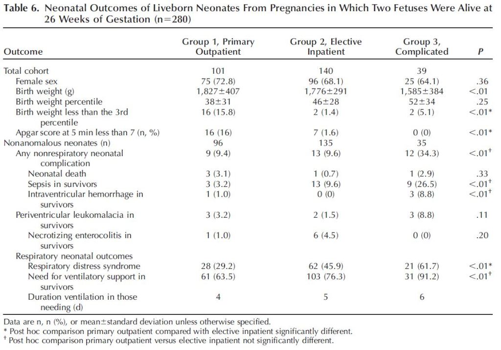
A more recent study with a greater sample size (193 momo gestations vs. 96) did not find a significant difference in outcomes between patients who went inpatient and those who went outpatient (Van Mieghem et al., 2014). The study differentiated between those who were outpatient from those who went electively inpatient, as well as from those who eventually went inpatient due to complications. The authors found that outpatients delivered at a slightly later gestational age (Table 1). However, more low APGAR scores were recorded in babies born from moms who went outpatient (Table 2). In the end, the authors concluded that they did not have enough statistical power to say whether one should be monitored as inpatient or outpatient. They did advise that the monitoring be frequent. According to the authors, the patients who were monitored as outpatient in this 2014 study were likely monitored more intensely than the outpatient cohort in the 2005 study. Intense outpatient monitoring in this case meant non stress testing (NST) four times a week and ultrasounds 1-2 times a week.

Figure 5. Table 5 from Van Mieghem et al., 2014 comparing pregnancy outcomes of patients who were monitored as outpatient (Group 1), inpatient (Group 2), and inpatient due to pre-existing complications (Group 3). Data show there was no significant difference in survival of twins delivered from moms who underwent outpatient monitoring vs. inpatient monitoring.
Figure 6. Table 6 from Van Mieghem et al., 2014, summarizing outcomes of liveborn newborns. Cohort of mothers who were monitored outpatient differed significantly from inpatient cohort in birth weight under the 3rd percentile (more babies under the 3rd percentile for weight in outpatient group), Apgar score (weaker neonates in outpatient group), and respiratory distress syndrome (fewer neonates with respiratory distress in outpatient group than inpatient).

Gestation beyond 34 weeks

Intense monitoring, whether inpatient or outpatient, begins around the age of viability, so between 24 and 28 weeks. But for how long should you expect to endure such tedious, albeit reassuring, surveillance? Assuming the monoamniotic pregnancy is not complicated by developmental anomalies, TTTS, or concerns from cord entanglement, the obstetric literature favors delivery between 31 and 34 weeks. Your OB will have his/her own expert opinion. In my own experience, my OB prefers delivering momos by 32 weeks, but is willing to accommodate to 34 if monitoring is increased to full-time. An influential paper (Roqué et al., 2003) recommended a 32 week delivery based on data from literature reviews that included 133 non-conjoined monoamniotic pregnancies. Data from that literature showed a significant increase in fetal mortality after 33 weeks, changing from 2-4% between 15-32 weeks to 11% from 33 -35 weeks. The percentage increased beyond 35 weeks (Figure). However, the study was criticized by later researchers, who reanalyzed the data and found that newborns delivered at 32 weeks died from fatal anomalies and so cautioned against such an early delivery (Dias et al., 2010). However, subsequent studies still support delivery before 34 weeks. The influential paper by Van Mieghem et al., 2014, discussed in the section above, recommends delivery of momo twins by 33 weeks. Figure 7 illustrates the interchange between the risk of prematurity and the threat of sharing an amniotic sac, based on their data. After 33 weeks, the risk of being in the womb outweighs any benefits.

Figure 7. Figure 1 from Van Mieghem et al., 2014 depicting interchange between risk of prematurity and risk of sharing an amniotic sac. The authors ultimately favor delivery at 33 weeks based on results from their data analysis.

Since 2014, different studies support different gestational ages for delivery. One retrospective study from 2015 supported delivery (including vaginal delivery!) within the 36th week, so long as there were no pregnancy complications (Anselem et al., 2015). Likewise, no babies died after delivery beyond 34 weeks (up to 36 weeks and 6 days) in a subsequent retrospective study (The MONOMONO Working Group, 2018). However, the authors of that study cautioned that their sample size of 10 twin sets delivered in the 36th week of gestation was too small to confidently recommend for future patients. Instead, the authors recommended the conventional 32 – 34 weeks and 6 days gestational age for delivery.

When considering Quality Adjusted Life Years (QALY), or the quality of life outcome from being born premature vs. risk of dying in the womb, another recent study supported an even earlier delivery time of 31 weeks (Hickock et al., 2018). The lowest rates of dying in the womb are associated with delivery at 28 weeks, while the lowest rates of newborn death and cerebral palsy occurred at 34 weeks. Thirty-one weeks was that optimal delivery time between the two outcomes (Fig. 8). However, when considering the exorbitant cost of a premature neonate to the families, the authors favored delivery at 32 weeks or even 33 weeks.

Figure 8. Graph from poster by Hickock et al., 2018, depicting quality of life outcomes for monoamniotic twins delivered at different gestational ages between 28 and 34 weeks. By their measures, 31 weeks gestational age is the optimal time for delivery (green line with stars plotted above). However, 32 and 33 weeks gestational age are preferred if one chooses to minimize hospital costs to families, since delivery at an earlier gestational age would coincide with a longer hospital stay.

Caesarean vs. vaginal birth

Many, if not most, women prefer to deliver their babies vaginally. So a mother who is diagnosed with monoamniotic twins may be disappointed that a caesarean delivery is in her future. While some hospitals and physicians will facilitate a vaginal delivery for momos, ACOG (American College of Obstetricians and Gynecologists) recommends that momos be delivered by caesarean section. Therefore, your hospital and physician likely will not accommodate your desire for a vaginal delivery. And you may want to resist the temptation to combat them on the subject. Afterall, vaginal birth of monoamniotic twins is risky. Caesarean delivery avoids risk of cord prolapse, where the cord of the second twin falls out of the cervix before the baby and can become clamped (Lewi, 2010).

Despite its risks, monoamniotic twins have been successfully delivered vaginally, and recent publications support vaginal delivery under optimal circumstances. For instance, vaginal delivery was deemed possible according to one study under the conditions that there were no preexisting complications, Twin 1 was head-down, and Twin 2 was not at least 30% greater in size than Twin 1 (Anselem et al., 2015). Another study published the following year compared the outcomes of 29 momo twin pairs (Khandelwal et al., 2016). Around half were delivered via c-section, and the other half were delivered vaginally. Nearly all had tangled cords at birth. All twin pairs were delivered around the same time (around 33 weeks gestational age). Survival outcomes were similar between twins born from caesarean section and the birth canal. However, the authors noted that the rate of intracranial hemorrhaging (uncontrolled bleeding in the head) was significantly lower in neonates born vaginally. Further, a nearly significant lower length of stay in the hospital and respiratory complications were correlated with that population of neonates. The authors conclude that vaginal delivery is a viable alternative delivery method for momos, although the sample size is small and is counter to the recommendation of the ACOG.

A much larger retrospective study in France compared 221 momo pregnancies (de Vergie et al., 2019). Patients were monitored both inpatient and outpatient with no significant difference in fetal mortality between the two groups. The average gestational age at time of delivery was 33-34 weeks. Of the patients who delivered at 34 weeks and beyond, one third delivered vaginally. Within the study cohort, there were not many differences in the outcomes of momo twins delivered vaginally or via c-section, except that the latter group was associated with lower Apgar scores and higher NICU submissions, once again suggesting the benefit of a vaginal delivery over caesarean section when possible.

Conclusions

If you are pregnant with monoamniotic twins in the United States, anticipate either going inpatient to the hospital, or visiting the perinatologists multiple times a week, beginning around the age of viability (26 to 28 weeks). At this time, the babies’ heartrates will be monitored with echocardiographs, and their growths will be measured from ultrasounds. Your intensive monitoring schedule will persist for the duration of the pregnancy until delivery around 32 weeks gestational age. If no risks to your babies are detectable, your physician may oblige you to let your twins gestate beyond 32 weeks in exchange for more frequent monitoring. Delivery will be via caesarian section.

Due to the rarity of this twin type, management of monoamniotic pregnancies is formed around theoretical risks. New studies suggest that some of the standard practices for managing monoamniotic gestation (e.g., inpatient monitoring, gestation up to 32 weeks, and caesarean delivery) are unnecessarily risky to the mother and babies’ quality of life. However, studies investigating monoamniotic twin pregnancy management often battle small sample sizes or are retrospective, which is problematic because one cannot test for causal relationships.

Best practices for managing monoamniotic twin pregnancy will change over time. The current protocol, which requires a demanding monitoring schedule and premature delivery by caesarean section, may not be your ideal birth plan. But it is correlated with a drastic rate increase in momo twin survival. Until more momos are born under a variety of circumstances to contribute data to future studies, be conservative and follow your physician’s advice. And breathe a sigh of relief because you and your babies are (likely) going to be okay!

References

Anselem, O., Mephon, A., Le Ray, C., Marcellin, L., Cabrol, D., and Goffinet, F. 2015. Continued pregnancy and vaginal delivery after 32 weeks of gestation for monoamniotic twins. European Journal of Obstetrics & Gynecology and Reproductive Biology 194: 194-198.

de Vergie, L., Dochez, V., Lorton, F., Riteau, A., Dumas, L., Riethmuller, D., Goffinet, F., Rozenberg, P., Thubert, T., Flamant, C., Arthuis, C., and Winer, N. 2019. 358: Management of monoamniotic twin pregnancies: Retrospective multicenter study of 221 cases. American Journal of Obstetrics and Gynecology 220: S247-S248.

Dias, T., Mahsud‐Dornan, S., Bhide, A., Papageorghiou, A.T., and Thilaganathan, B. 2010. Cord entanglement and perinatal outcome in monoamniotic twin pregnancies. Ultrasound in Obstetrics and Gynecology: The Official Journal of the International Society of Ultrasound in Obstetrics and Gynecology 35: 201-204.

Hack, K.E., Derks, J.B., Schaap, A.H., Lopriore, E., Elias, S.G., Arabin, B., Eggink, A.J., Sollie, K.M., Mol, B.W.J., Duvekot, H.J., Willekes, C., Go, A.T., Koopman-Esseboom, C., Vandenbussche, F.P., and Visser, G.H. 2009. Perinatal outcome of monoamniotic twin pregnancies. Obstetrics & Gynecology 113: 353-360.

Heyborne, K.D., Porreco, R.P., Garite, T.J., Phair, K., and Abril, D. 2005. Improved perinatal survival of monoamniotic twins with intensive inpatient monitoring. American journal of obstetrics and gynecology 192: 96-101.

Hickok, R.A., Walker, A.R., and Caughey, A.B. 2018. 236: When to deliver monochorionic-monoamniotic twins undergoing inpatient continuous fetal monitoring–A decision analysis. American Journal of Obstetrics & Gynecology 218: S153-S154.

Khandelwal, M., Revanasiddappa, V.B., Moreno, S.C., Simpkins, G., Weiner, S., and Westover, T. 2016. Monoamniotic Monochorionic Twins—Can They Be Delivered Safely Via Vaginal Route? Obstetrics & Gynecology 127: 3S.

Lewi, L. 2010. Cord entanglement in monoamniotic twins: does it really matter? Ultrasound in obstetrics & gynecology: the official journal of the International Society of Ultrasound in Obstetrics and Gynecology 35: 139-41.

MONOMONO Working Group. 2018. Inpatient vs outpatient management and timing of delivery of uncomplicated monochorionic monoamniotic twin pregnancy: the MONOMONO study. Ultrasound in obstetrics & gynecology 53: 175-183.

Myrianthopoulos, N.C. 1975. Congenital malformations in twins: epidemiologic survey. Birth Defects Original Article Series 11: 1-39.

Roqué, H., Gillen-Goldstein, J., Funai, E., Young, B.K., and Lockwood, C.J. 2003. Perinatal outcomes in monoamniotic gestations. The Journal of Maternal-Fetal & Neonatal Medicine 13: 414-421.

Rossi, A.C., and Prefumo, F. 2013. Impact of cord entanglement on perinatal outcome of monoamniotic twins: a systematic review of the literature. Ultrasound in Obstetrics & Gynecology 41: 131-135.

Tongsong, T., and Chanprapaph, P. 1999. Evolution of umbilical cord entanglement in monoamniotic twins. Ultrasound in Obstetrics and Gynecology: The Official Journal of the International Society of Ultrasound in Obstetrics and Gynecology 14: 75-77.

Umur, A., van Gemert, M.J., and Nikkels, P.G. 2003. Monoamniotic-versus diamniotic-monochorionic twin placentas: anastomoses and twin-twin transfusion syndrome. American journal of obstetrics and gynecology 189: 1325-1329.

Van Mieghem, T., De Heus, R., Lewi, L., Klaritsch, P., Kollmann, M., Baud, D., Vial, Y., Shah, P.S., Ranzini, A.C., Mason, L., Raio, L., Lachat, R., Barrett, J., Khorsand, V., Windrim, R., and Ryan, G. 2014. Prenatal management of monoamniotic twin pregnancies. Obstetrics & Gynecology 124: 498-506.

Feel free to share your monoamniotic pregnancy game plan in the comments below!

Diagnosing my momo pregnancy

Learning I have monoamniotic twins

Expecting twins adds an element of risk to the pregnancy. The mother is at greater risk of anemia, gestational diabetes, preeclampsia, and preterm labor, to name a few complications. The fetuses are at risk of complications arising from premature birth. The chance of the mother having to deliver via Cesarean section increases with having multiples. I had to come to terms with all of this as soon as I learned I had twins. When my blood screen results confirmed that my twins were identical, I had more risks to add to my worry list. Monochorionic monozygotic twins have more complications than dichorionic twins. Because most monozygotic twins are also monochorionic, I had to accept that now my twins were also at risk of structural anomalies which could affect one or both or their hearts, or were at risk of twin-to-twin transfusion syndrome (TTTS). If, God forbid, a twin were to die in utero, then the chance of the other twin dying from acute intertwin transfusion increases for monochorionic twins because of the anastomoses of blood vessels in the placenta of monochorionic twins. In the occurrence of acute intertwin transfusion, a drop in blood pressure from the demised twin will cause the blood to suddenly backflow from the surviving twin, potentially fatally damaging vital organs, or at least damaging the brain. And, on the off-chance that the girls were monoamniotic, I could add sudden death from umbilical cord constriction to my list of complications to worry about.

In the two weeks between my OB appointment and my next ultrasound with the Maternal-Fetal Medicine (MFM) specialist, my mind was plagued with doubt as to whether or not my girls were still alive. Perhaps I sound overly pessimistic, but experiencing multiple losses will do that to a person. I arrived at my perinatal appointment on pins and needles. But I could finally breathe a sigh of relief when the ultrasound tech at the perinatology clinic confirmed both twins were alive and well. I was elated. The lightest I’d felt in days. Then the MFM doctor arrived.

He scrutinized the ultrasound. The twins were clearly monochorionic; he was looking for the intervening membrane to confirm they were diamniotic.

“I’m not seeing an intervening membrane,” he told the tech. The mood in the room suddenly changed. The smile on my face flipped a 180. Everyone was silent, intently studying the ultrasound screen. He turned his attention to the twins’ umbilical cords. Doppler imaging highlighted their cords in an unambiguous twist. Their insertions onto the placenta were suspiciously close to one another. The absent intervening membrane and the close cord insertions were suggestive of a monoamniotic pregnancy (Fig. 1). The twisted umbilical cords hammered the nails into the proverbial coffin. In all my readings of twins, not much was mentioned of monoamniotic twin pregnancies other than they were marked by complication and high mortality. My rush of relief quickly re-subsided into gloom and anxiety. “I’m sorry,” the tech said to my husband and me as we left the room.

Figure 1. Ultrasound snapshots suggesting a monoamniotic pregnancy. Top: Looking at the top of the twins’ heads at 16 weeks 4 days gestational age. No intervening membrane is visible between the heads. Absence of evidence is not evidence of absence, but monoamniotic gestation cannot be ruled out based on the above image. Bottom: Umbilical cords are inserted close together on the placenta, indicated by arrows, which is much more common with monoamniotic identical twins than diamniotic (Zhao et al., 2014). Sadly, I do not save an ultrasound image of the tangled cords. Fetus is 18 weeks and 4 days gestational age in the bottom photo.

I would have to wait a grueling two weeks before I could talk about my momo diagnosis with my OB. It was the most unbearable two weeks of my life. Time passed impossibly slow. Not even the anticipation of Christmas Day as a child felt so painful. All I could do to stay sane was research the answers to all of my nagging questions. What was the prognosis of the twins I was carrying? What made monoamniotic pregnancies so risky? How would this diagnosis change my prenatal care? The research process would take me on an emotional roller coaster ride, but the final impression was that we were going to make it off the ride alive.

My next post will summarize the takeaways I gathered for what to expect on my momo pregnancy journey. It was the inspiration for starting my blog.

Feel free to share your experience of discovering your twins were monoamniotic in the comments below!

What are twins, anyway?

Di-di, mono-di, mono-mono. I heard these terms for the first time from my OB as soon as I learned I was having twins. After my OB threw a bunch of twin jargon at me, I knew it was time to begin my homework. What were these other types of twins that I did not know about? Why were some types more risky to carry than others? What was I to expect during this twin pregnancy?

Fraternal twins
Identical twins: di-di and mo-di
Identical twins: mo-mo
Identifying twin type in early pregnancy

Fraternal twins

Broadly speaking, there are two types of twins: fraternal and identical. A mother can release two eggs simultaneously, and each one can be fertilized and implant itself into the endometrium of her uterus. Because these twins arise from two separately fertilized eggs, they are termed dizygotic (di-two, zygote=fertilized egg). They grow like any other embryo (Fig. 1). After about four days of cell division, each fertilized egg, or zygote, forms a hollow cavity and is called a blastocyst (Fig. 1A). The blastocyst contains an outer sphere of cells that will play a role in implantation, and an inner mass of cells that will develop into the embryo, its nutritive yolk sac, and its protective amnion. A week after fertilization, the zygote will have completely burrowed into the mother’s endometrium (Fig. 1B). In the endometrium, cells from the outer cell mass proliferate and will contribute to the chorionic membrane and placenta. The inner cell mass begins to differentiate into a dorsal mass and a ventral mass. Cavities form in the dorsal mass and the ventral mass. The ventral mass will develop into the yolk, which nourishes the embryo until the placenta is developed. The dorsal mass forms a cavity called the amniotic cavity, which will grow with the embryo and fill with fluid. The cells of the dorsal mass contacting the chorion will develop as the amniotic membrane while the cells contacting the yolk will develop as the embryo. Because two fertilized eggs have each buried themselves into the mother’s uterus, each baby develops its own chorion/placenta, and its own amnion (Fig. 2). Therefore, these twins are dichorionic-diamniotic or di-di. Dizygotic twins are as genetically similar to one another as any other sibling pair, and are known more colloquially as fraternal twins.

Figure 1. Anatomy of a zygote. (A) Cross section through blastocyst, a zygote that has formed a cavity surrounded by an outer cell mass and an inner cell mass. (B) Blastocyst implanted into the endometrium, or deciduous portion of uterus.
Figure 2. 40 day-old human embryo. In a di-di pregnancy, each embryo develops as a singleton embryo would. Here, the embryo is visible floating in its amniotic sac. The yolk sac is in the extracoelomic space within the chorion. The fuzzy-looking chorionic villi surround the entire surface of the chorion this early in development. They comprise the fetal portion of the placenta. Image credit: Ahokas and McKinney, 2008)

Identical twins: di-di and mo-di

The second type of twin, identical twins, is where twin classification becomes a little more complicated. They are termed identical because their DNA is nearly 100% identical. This happens when a mother releases one egg, as she ought to, but then after fertilization that egg throws a curve ball at her and splits itself into two zygotes! Because these two zygotes began as one, identical twins are also called monozygotic (mono-one, zygote=fertilized egg). Why would a zygote split? The cause is not yet well understood, but what is known is that for a brief moment during cell division, the tight connections between cells of the zygote lose integrity and the mass of future embryonic cells splits apart. If the split occurred very early, within the first three days after fertilization while the zygote is still aiming for the perfect real estate in the mother’s uterus, they will each develop their own chorion, placenta, and amnion (Fig. 3A). They will become, you guessed it, di-di twins. Just like fraternal twins! Only, remember, these twins have 100% of each other’s DNA in common. If this zygote splits into two after it has buried itself into the endometrium and developed its chorion, but before it has developed its amnion, then each embryo will share the chorion and placenta, but dwell in their own amniotic cavity. This schism would have to occur between 4 and 7 days after fertilization (Fig. 3B) and these twins would be referred to as mono-di or mo-di.

Figure 3. Timeline of a zygote. (A) Cell division, or mitosis, of the fertilized egg occurs approximately 30 hours after fertilization. A complete division of this zygote will result in two identical dichorionic-diamniotic twins. (B) Between four and seven days of cell division, a zygote is in the body of the uterus and may begin implanting into the endometrium. This is when the future placenta begins to develop. If the zygote splits at this point, the resulting twins will share a placenta but each embryo will develop its own amnion, classifying them as monochorionic-diamniotic twins. (C) After 7 days of mitosis, the zygote has established its chorion and begun to develop its amniotic cavity and membrane. A split in the embryonic mass (see Fig. 1) at this point will result in identical twins who share a placenta and an amniotic sac, classifying them as monochorionic-monoamniotic.

Identical twins: mo-mo

Identical twins are pretty rare. Mo-di identical twins are actually more common than di-di identicals, despite the fact that fraternal twins (which are always di-di) are the most common type of twin overall. But the most uncommon twins of all, the rarest of the rare, are the monochorionic-monoamniotic twins, a.k.a. monoamniotic, mono-mono, and momos. They occur in 1% of all twin pregnancies. That’s about 1 in 60,000 pregnancies! These twins are susceptible to a host of problems including developmental anomalies and cord compression, along with other problems that come with having mo-di twins such as Twin-to-Twin Transfusion syndrome or TTTS.

Momo twins result from the zygote splitting after the 8th day post-fertilization (Fig. 3C). The unfortunate circumstance of splitting so late is that one of the zygotes may not acquire the proper unspecialized cells it needs to completely grow itself. Incompletely developed fetuses or midline developmental defects are more common in monoamniotic pregnancies. One could imagine that the variety of developmental issues common in momos lies on a continuum based on the timing of separation. A very late split of the zygote will result in conjoined twins, who may share a heart or part of the GI system. Sadly, such twins seldom survive long after birth, if they make it that far.

Identifying my twin type in the early first trimester

Of course as soon as I learned I was having twins, I was aching to know which type I was carrying. And like any other pregnant woman, I wanted to know their biological sex. Statistically, I was going to have boy-girl fraternal twins. Fraternal twins are 66% more common than identical twins. Further, my demographic would suggest they are likely fraternal since fraternal twins more commonly occur in non-Hispanic whites.

However, unlike identical twins, fraternal twins typically run in families. Fraternal twins do not run in my family. It is of no consequence to me if they run in my husband’s family since he does not affect how many eggs I release (sorry hubs, I know you like to imagine your extreme manliness can cause twins). But for the record they do not run in his family either. Therefore, my twins were a freak occurrence and quite possibly identical. Further, my ultrasounds did not look like early ultrasounds of dichorionic twins (Fig. 4A). Even though the membranes are thin at eight weeks, there is often a characteristic mountain peak or lambda shape visible where the placentae abut one another (Fig. 4B). On mine, there was no peak (Fig. 4D). Sharing a placenta would have to mean my twins were identical. So then I had a 50:50 chance of carrying boys or girls. Unless they were monoamniotic; then my chances of having two girls would double since twice the number of momos are female than male. But monoamniotic is not very likely. Could my babies be one in 60,000? No way.

Oi. I couldn’t wait for the 20-week ultrasound to answer these questions. I needed a blood test.

Figure 4. First trimester ultrasounds (US) of twins. (A) Early first trimester US of di-di twins, well separated in their own gestational sacs with two yolk sacs visible. (B) Late first trimester US of di-di twins when characteristic lambda sign (arrow) is visible. Lambda formed by thick, four-layer membrane at its placental insertion. (C) Late first trimester US of mono-di identical twins with diagnostic T-sign (arrows), formed by thin, two-layer intervening membrane attaching to shared placenta. (D) 8w4d US of my monoamniotic twins. At this stage, diagnosis of amnionicity is tentative. But note the absence of a thick dividing membrane (putatively ruling out dichorionicity) and single yolk sac (indirect indicator of monoamnionicity, Bromley and Benacerrof, 1995). Image credit: A-C from Fuchs and D’Alton, 2018

References

Ahokas, R.A. and McKinney, E.T. 2008. Development and physiology of the placenta and membranes. The Global Library of Women’s Medicine. ISSN: 1756-2228) 2008; DOI 10.3843/GLOWM.10101

Bromley, B. and Benecerrof, B. 1995. Using the number of yolk sacs to determine amnionicity in early first trimester monochorionic twins. Journal of Ultrasound in Medicine 14: 415-419.

Fuchs, K.M. and D’Alton, M.E. 2018. Monochorionic monoamniotic twin gestations In J.A. Copel, M.E. D’Alton, H. Feltovich, E. Gratacós, D. Krakow, A.O. Odibo, L.D. Platt, and B. Tutschek (Eds.), Obstetric Imaging: Fetal Diagnosis and Care (pp. 642-645.e1). Elsevier, Inc. Retrieved from https://doi.org/10.1016/B978-0-323-44548-1.00159-5

———————– 2018. Monochorionic diamniotic twin gestations In J.A. Copel, M.E. D’Alton, H. Feltovich, E. Gratacós, D. Krakow, A.O. Odibo, L.D. Platt, and B. Tutschek (Eds.), Obstetric Imaging: Fetal Diagnosis and Care (pp. 645-648.e1). Elsevier, Inc. Retrieved from https://doi.org/10.1016/B978-0-323-44548-1.00160-1

———————– 2018. Monochorionic monoamniotic twin gestations In J.A. Copel, M.E. D’Alton, H. Feltovich, E. Gratacós, D. Krakow, A.O. Odibo, L.D. Platt, and B. Tutschek (Eds.), Obstetric Imaging: Fetal Diagnosis and Care (pp. 648-650.e1). Elsevier, Inc. Retrieved from https://doi.org/10.1016/B978-0-323-44548-1.00161-3

The Journey Begins

You have twins! Or: Is more really merrier?

The discovery
Early symptoms of a twin pregnancy
My momo journey

The discovery

After multiple losses and seven months of actively trying to conceive a sibling for my daughter, I was pretty anxious about my first OB appointment proceeding my latest positive pregnancy test. Six months earlier, I was in the same clinic anticipating my first glimpse of the 9.5 week old embryo inside of me. When my physician fired up the ultrasound, I saw a tiny bean on the screen. It looked far tinier than I remembered my daughter looking when she was an 8 week embryo. Then my OB measured the heartbeat. It was slow, and the embryo measured three weeks behind. My baby was dying. Though I knew that those first few weeks of pregnancy are the most tenuous, the memory of the pain I endured from my last chemical pregnancy took over me and I nearly fainted in front of my OB and husband. Now I was back in the same room, this time without my husband, about to confirm a pregnancy I was hoping for over two years prior.

The profile of an embryo flashed on the monitor. It looked much larger than the last poor embryo I lost. That was a comforting sight! “Looks good,” my physician assured me as she continued to survey my pelvic cavity with the ultrasound wand. The baby slipped from view but then suddenly re-emerged, this time in a frontal view. I was very confused as to how I could see a baby in profile and then almost instantly view it head on. “I don’t understand how to read ultrasounds,” I thought to myself. But my OB grew suspicious. She was silent for what felt like forever, studying the ultrasound monitor very intently. I began to feel slightly tense.

Suddenly, two embryos appeared on the screen (Fig. 1). She found what she was looking for.

“F**************k,” I groaned under my breath. I’m not usually one to swear.

Figure 1. Two embryos in a yin-yang orientation. The left twin (to the right in the image) is facing the viewer and its head is up. The right twin (to the left in the image) is in profile and its head is down. The gestational age of the embryos is 8 weeks and 4 days.

I began to panic. I couldn’t believe this was real. Twins don’t run in my family, how could this happen to me? I have read some women’s reactions to the news that they are bearing twins. Some women have wanted twins all their life and are elated when they receive the news. Clearly, from my reaction, I was not one of those women. Some people look at parents juggling twin babies and feel pity for them, unable to fathom the exorbitant amount of work it must take to raise more than one child simultaneously. This was not crossing my mind, either. I knew very little about twins, but what I did know at the time of the diagnosis was that carrying twins comes with a host of difficulties and immediately launches my pregnancy into high-risk. After suffering through losses, and waiting years to make another baby, I have made two babies with a tenuous outcome. Instead of dealing with an early loss of one baby, I could face a late loss of two babies. How could I possibly endure it?

“I’ve heard of cases with twins where one dies very early and gets absorbed by the other. Is that still a possibility?” I foolishly asked my OB in hope that my situation might turn into a normal singleton pregnancy.

“Not at this point,” she assured me. The embryos were too far along and looked too healthy, which of course was actually a good thing. She continued, “Now we’ll need to figure out if they’re di-di, mono-di, or mono-mono.” These words were completely foreign to me. I knew twins came in two flavors: fraternal (dizygotic) and identical (monozygotic). “If they are mono-mono then we have a problem but those are extremely rare. These are likely mono-di or di-di. Di-di is the safest, but we can work with mono-di. Right now the membranes are too thin to tell what type of twin you have, but we’ll be able to tell over time when the membranes get thicker.”

My head was spinning. I was sent home with a packet of standard pregnancy pamphlets, plus a giant list of supplements to take especially for women expecting twins.

My husband could not attend the appointment because he had a class to teach that morning. I got out of the appointment 15 minutes before his class would begin so I gave him a call.

“I have good news and bad news,” I told him. “Good news is the baby looks healthy! Bad news, if you want to call it that, is there are actually two babies. And with that, I’m going to let you go so you can teach your class.” My husband began laughing. I could tell he was smiling ear to ear. I wasn’t sure how he would handle the news but I wasn’t expecting him to be so happy about it. I’ve got to say that his enthusiasm did ease my mind a bit, and for the first time since the initial discovery, I began to feel a little excited too.

Early symptoms of a twin pregnancy

After my twin diagnosis, a lot of my early pregnancy symptoms began to make sense. My symptoms had surprised me for being stronger than I remember with my first, singleton pregnancy. In retrospect, five early symptoms were possibly indicative of twins.

1. A dark line on my pregnancy test. I could not wait to find out if I was having another baby so I did the one thing I told myself not to do: I peed on the stick early. Only 10 days after ovulation. Yes I was using a first response early detection test, but results are not guaranteed so early. Yet there it was, an unambiguous positive staring back at me (Fig. 2). I texted the pic to a friend. “Wow! That looks dark for only 10 days,” was her response. In hindsight, yes, I suppose it was.

Figure 2. Home pregnancy test, early response test, taken just 10 days past ovulation. Arrows indicate test line. The line looked darker in person, I swear! 😊

2. Extreme fatigue. How extreme? Here’s an example: My daughter (Fig. 3) was going through her “tattoo artist” phase. She would regularly give me a tattoo at 7:30 or 8 p.m. Using my expensive gel pens, she would scribble all over my hand. The metal tips of the pens would dig into my flesh; it’s a wonder I didn’t bleed. She would merrily inquire, “Does that tickle mommy?” But I couldn’t answer her. I was already passed out on the bed…at 7:30…while pen tips scraped off my skin.

Figure 3. Beatrix, the Elder. Do not let her innocent face fool you. She was more than willing to take advantage of my condition to inflict her masochistic urges upon me.

3. Insatiable hunger. Anyone who knows me knows I love food. But my hunger in early pregnancy felt pathological. Every minute of the day, I was thinking about food. I was fantasizing about my next meal while I was still eating my current meal! And when I was so full I wanted to vomit, I still thought about what I could eat next. Sometimes I was afraid of my own appetite.

4. Intense nausea and vomiting. Speaking of vomiting. Yeah. This symptom is a little strange for me. Some women experience debilitating nausea during their first trimester. I’m not one of those women. I felt nauseous when I was expecting my daughter, but it was on and off and quite manageable. I may have vomited once. And with this pregnancy, I told my husband repeatedly that I felt less nauseous than with my first. My vomiting record says otherwise. Though I didn’t feel an omnipresent nausea, I vomited almost daily (sometimes twice daily) when brushing my teeth. I am in my second trimester and I still vomit. Further, buffalo plaid was coming into season at the height of my morning sickness, and I found that just looking at someone wearing the red-and-black pattern would start me dry heaving. Maybe I was more nauseous than I wanted to admit.

5. Child psychic abilities. Okay, this one has no scientific merit. But sometimes people swear their child knows when they are pregnant, or knows the gender before the parents know. Once my husband and I knew we were expecting, we asked our daughter if she was excited for a baby brother or sister. She told us she was very excited for her baby brother AND sister. “You’re only having one,” we warned her. “No, we’re having two,” she would laugh in response. Really, she was warning us.

My momo journey

After the twin diagnosis, my pregnancy experience would be vastly different from my previous singleton pregnancy. Because my pregnancy was now classified as high-risk, I would have to visit a maternal fetal medicine (MFM) specialist at the perinatology clinic every two weeks. The upside would be plenty of ultrasounds. As long as the lower of the two twins was head-down by 24 or so weeks, I could anticipate a vaginal delivery as late as 37 weeks. The book, When You’re Expecting Twins, Triplets, or Quads, by Dr. Barbara Luke et al. (Fig. 4) quickly assured me that a twin pregnancy did not have to result in underweight, fatally premature, or developmentally malformed offspring. I highly recommend the book to anyone expecting multiples. Despite my optimism, a later surprise diagnosis would launch my pregnancy into extreme lows but also some highs, and I would have a lot more to learn about what it takes to safely carry twins. I don’t know where my journey through twin pregnancy will take me. But I hope by the end I will be taking home two healthy momo twins. Please join me on my momo journey!

Figure 4. “When You’re Expecting Twins, Triplets, or Quads,” by Dr. Barbara Luke et al. A must-read for anyone carrying more than one fetus.

Please feel free to share your experience of when you learned you were expecting multiples in the comments below!